The retina, located in the back of the eye, is an essential layer of cells where light is detected and sends a signal to your brain via photoreceptors. These signals allow you to understand and process the images around you. The retina, albeit small, plays a major role in vision. However, it is prone to various conditions and diseases, such as retinal tears and retinal detachments, that can lead to vision loss if left untreated.
Retinal Tear
Retinal tears typically involve the vitreous gel adhering to the retina and pulling on it over time. If tugging is persistent or strong enough, it will cause a tear. These tears can allow liquid vitreous to get under the retina. This could lead to a retinal detachment. Tears are relatively common, especially in patients over the age of 50 and those with myopia (near-sightedness) or a family history of retinal detachments.
Retinal Tear Symptoms:
- Sudden or an increased amount of floaters
- Black spots in vision
- Blurred or darker vision
- Loss of peripheral vision
- Flashes of light
Retinal Tear Treatments:
- Laser surgery (photocoagulation): Pulses of laser energy are directed to specific spots around the retinal tear. A retina surgeon then “welds” the torn retina to the underlying tissue to prevent further tearing.
- Freezing (cryopexy): An extremely cold surgical probe freezes areas around the retinal tear. Once a local anesthetic is applied to the eye, the probe is applied to the eye’s outer surface, creating a scar that secures the retina to the wall of the eye.
Retinal Detachment
Retinal detachments are more severe than retinal tears. This signifies that the retina has separated from the back of the eye. Detachments are usually caused by fluid getting underneath the retina through an untreated retinal tear.
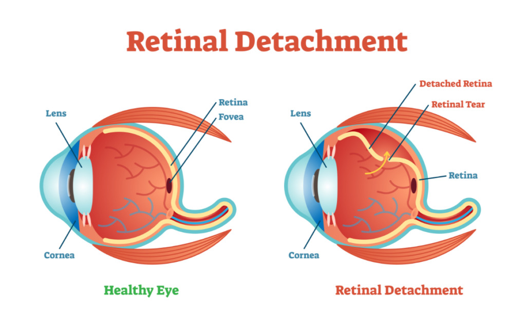
There are three types of retinal detachments: rhegmatogenous, tractional, and exudative. Rhegmatogenous retinal detachments are due to tears that enable fluid to get under the retina and detach it from the back of the eye. This is the most common and most dangerous as it happens quickly. Tractional retinal detachments are caused by scar tissue on the retina. This is less rapid than rhegmatogenous detachments and is typically seen in patients with diabetes or vascular occlusions. Exudative retinal detachments are caused by fluid leaking underneath the retina without tears. This is typically the result of other retinal diseases, injury to the eye, inflammation, or even cancers.
Retinal Detachment Symptoms:
Symptoms of a retinal detachment are initially similar to a tear (flashers, floaters, blurred vision). However, a patient might experience a dark shadow or “curtain effect” appearing over one side of their vision over time.
Retinal Detachment Treatments:
- Pneumatic Retinopexy: a retinal surgeon injects a gas bubble into the vitreous of the eye. The head is positioned so that the gas bubble floats to the detached area, which in turn presses against the detachment. The gas bubble flattens the retina until a seal forms between the retina and the eye wall. Cryopexy and photocoagulation are utilized to seal the tear. After about 1-3 weeks, the eye gradually absorbs the gas bubble and any extra fluid.
- Scleral Buckling: a synthetic band is attached to the outside of the eye, gently pushing the eye toward the wall near the detached retina. Over time, the retina becomes reattached to the eye wall.
- Vitrectomy: a retinal surgeon replaces the vitreous using gas injections. This gas pushes the retina against the eye’s wall and reattaches it. The eye naturally makes fluid that replaces the gas.
Sudden floaters, vision loss, or “shadows” in your vision are not typical. Untreated retinal tears or detachments can lead to irreversible vision loss. Should you have symptoms related to retinal tears or detachments, contact our office today at 954-772-3337. We will be glad to schedule a consultation with one of our retinal specialists, Dr. Burgess, Dr. Lara, and Dr. Villate.